Imaging
Optoacoustic (OA) Tomography produces images of the absorbed optical energy in acoustic sources generated in the tissue volume by nanosecond laser pulses. OAT combines advantages of optical illumination (high contrast and molecular specificity) and ultrasound detection (high resolution and depth of imaging)
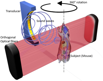
LOUIS-3D implements the optoacoustic technology for imaging the small animals for imaging the vasculature, organs, skeletal system and the skin. The volumetric images give us:
3D Mouse Optoacoustic Imaging System
The system demonstrated its capability to display quantitatively accurate contrast of vasculature, bones and various organs based on variations in blood content. When combined with targeted contrast agents, LOIS-3D images of tissue can be obtained with molecular specificity. Thus, LOUIS-3D system is useful in biomedical research involving animal models.
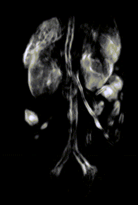
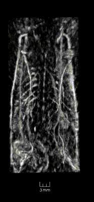
Functional Imaging
Functional optoacoustic provides a measurement of [Hb] and [HbO] (hematocrit) in tissues and blood vessels, assessment of heart function and blood flow. It allows for assessment of tumor angiogenesis, angiography, detection and characterization of stroke and traumatic injury of the brain.
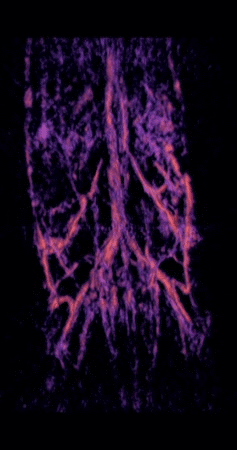
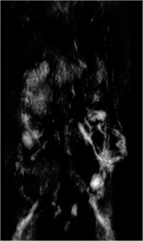
Arterial system of a mouse at 1064nm/757nm
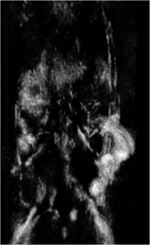
Venous system of a mouse at 1064nm/757nm
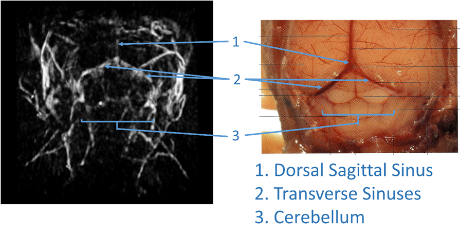
Optoacoustic imaging of a live mouse brain, total hemoglobin map
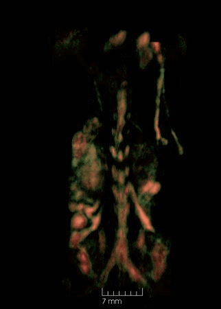
Molecular Imaging
Molecular optoacoustic imaging can show distribution of cellular receptors with targeted nanoparticle agents. It is also useful for studies into kinetic and dynamic distribution of contrast agents.
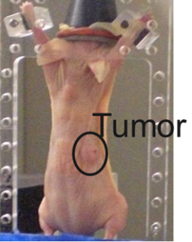
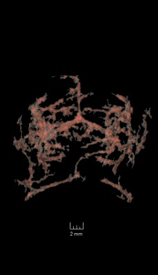
The images to the right are molecular optoacoustic images of the tumor seen in the picture above.
Angio-Endogenous
GNR-PEG-Herceptin
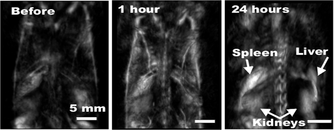
Image 2D Projections: Mouse Injected with Contrast Agent
After intravenous injection of PEGylated gold nanorods as contrast agent, microvasculature as small as 50 micron in soft tissue and even bones (ribs and spine) was visualized even though the system spatial resolution is only 500 micron. Redistribution kinetics of gold nanorods (GNR) was observed in tissue showing presence of GNR in the vasculature shortly after injection, then gradual transfer of the contrast to the organs (such as liver and spleen) serving as filters for nanoparticles.