Problem and Solution for Breast Cancer Imaging
(X-ray Mammography vs. US vs. OAT)
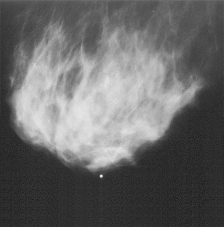
MAMMOGRAPHY IMAGE
Missing >45% of cancerous tumors due to low contrast in dense breast
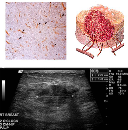
ULTRASOUND IMAGE
Medium Contrast
Resolution: 0.5 mm
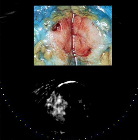
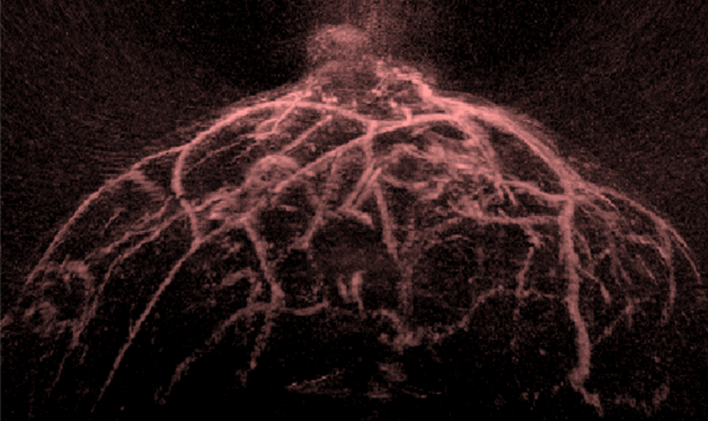
OPTOACOUSTIC IMAGE
High Contrast: 2x-3x
Resolution: 0.5 mm
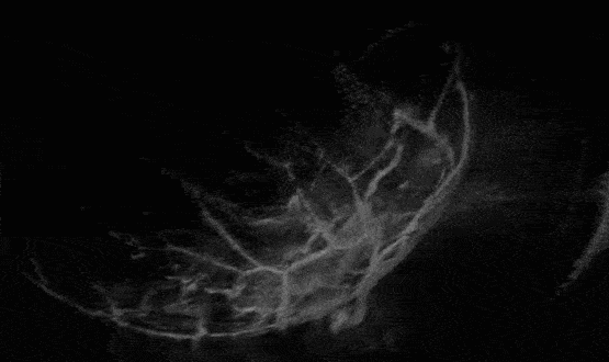
LOUISA-3D Imaging System for Breast Cancer Research
(click to see animation)
LOUISA-3D is a non-invasive clinical breast imaging that produces three-dimensional volumetric images of the breast, along with two-dimensional ultrasonic images for image co-registration.
The video shows the working of LOUISA-3D breast imaging system. This non-invasive breast imaging system produces images based on optoacoustic technology. Within minutes, it produces scans at different wavelengths for visualization of different features of the tissue, and causes negligible discomfort to the patient. The system come with an in-house software program that processes the volumetric data as per the user requirements, such as, visualization of vasculature, skin, tumors or quantitative information such as oxygen saturation and hemoglobin content.