In recent years, motivated by the research community to obtain innovative solutions, biomedical imaging has made remarkable strides, thanks in large part to innovations in the realm of optoacoustic (photoacoustic) systems and high-end ultrasound. From the emergence of cutting-edge quantitative 3D full-view optoacoustic (photoacoustic) tomography systems to the development of transmission/reflection ultrasound tomography systems for biomedical applications, the landscape of biomedical imaging research is more vibrant than ever. But why, specifically, should biomedical imaging centers, small animal research laboratories, and pharmaceutical companies, especially the ones in the bustling biomedical research landscape, prioritize upgrading to this latest technology?
A Closer Look at the Tech
At its core, optoacoustic (photoacoustic) imaging technology combines the principles of optical imaging where light is absorbed by molecules, with ultrasound as a means to deliver imaging information for processing by computer. When near-infrared light interacts with biological tissues, being absorbed by molecules, it produces a small amount of heat and, in turn, generates an ultrasound signal through thermal expansion, providing images that are molecular and (unlike pure optical images) are highly resolved details of molecular composition. Ultrasound imaging, on the other hand, is mechanical, it is based on reflections of ultrasound waves from tissue boundaries due to differences in density or transmissions of ultrasonic waves through tissues at different speeds of sound. Thus, optoacoustic (photoacoustic) and ultrasound images provide complementary information. Essentially, dual-modality systems combining optoacoustic (photoacoustic) plus ultrasound images merge the best of both worlds, giving unprecedented insights into the molecular composition within details of anatomical tissue structures. When nanoparticles with strong optical absorption properties are used as exogenous targeting contrast agents, researchers have an extraordinary opportunity to visualize not only the distribution of hemoglobin and oxyhemoglobin as the dominant optical chromophores in live tissues, but also visualize proteins that do not absorb near-infrared light, but play important role in tissue functions or development of abnormal conditions, such as cancer.
Comparison with conventional imaging technologies
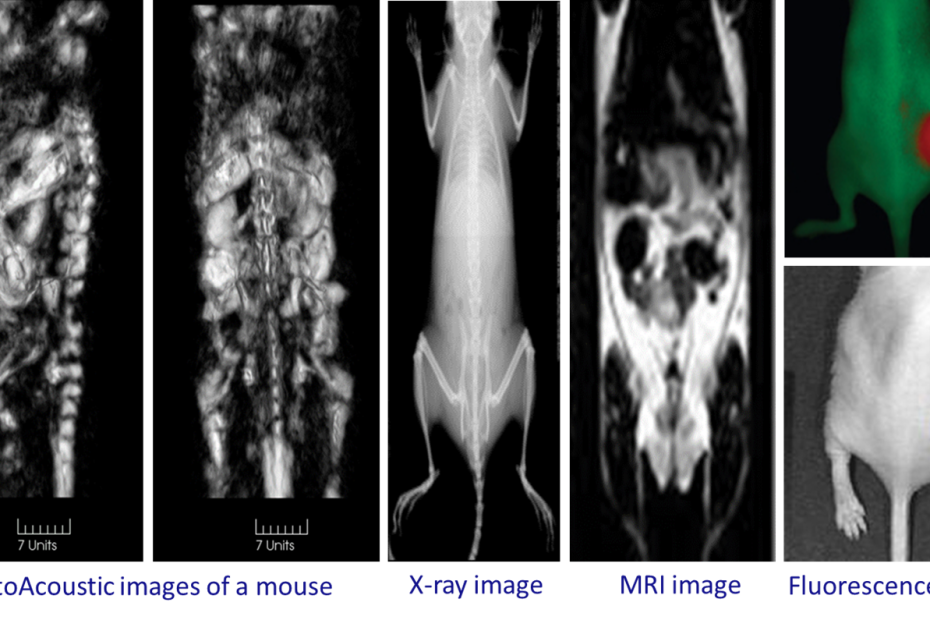
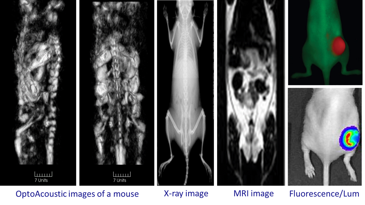
Let us discuss the advantages of the optoacoustic (photoacoustic) imaging dual modality combined with ultrasound by comparing its features and specifications with conventional imaging technologies used in research:
X-ray CT. Soft (water-containing) biological tissues are relatively transparent for X-rays but hard tissues such as bones are opaque for X-rays. Thus, x-ray images display skeleton and calcifications with excellent contrast and resolution, however, X-ray-based contrast in various soft tissues is low. Optoacoustic (photoacoustic) images are based on optical absorption of red blood cells and provide images of bones and other live hard tissues based on the concentration of microvasculature. Ultrasound can provide images of bones and hard tissues based on strong reflections from their boundaries. Thus, optoacoustic plus ultrasound dual modality can in many applications replace X-ray CT while additionally providing high-contrast imaging of soft tissues.
Fluorescence Imaging Systems. Fluorescence is based on light absorption by molecules and thus provides advantages of molecular contrast similar to optoacoustic (photoacoustic) imaging. However, fluorescence light re-emitted by molecules is strongly scattered through biological tissues before reaching detectors of the imaging system. As a result, fluorescence imaging has poor spatial resolution and associated lost sensitivity to optical contrast in the depth of biological tissue. Optoacoustic (photoacoustic) imaging provides high molecular tissue contrast and excellent spatial resolution in the depth of biological tissue due to the fact the information is delivered to the detectors by acoustic waves that experience negligible scattering compared with strong optical scattering.
Magnetic Resonance Imaging. MRI is a universal imaging technology that provides high-contrast images of both hard and soft tissues based on the density of hydrogen atoms (water and lipids) and the environment in which hydrogen atoms interact with surrounding atoms of molecules composing tissue structures. MRI is the most versatile imaging technology applicable to all different tissues not limited to depth from the surface. However, MRI can match neither the capabilities of optoacoustics (photoacoustics) for functional imaging of blood oxygen saturation and total hemoglobin concentration nor its capabilities for highly specific molecular imaging where the selectivity to molecules or nanoparticles is provided by the tunable laser wavelength. Furthermore, the high spatial resolution of MRI is provided by magnets with high magnetic fields that are very expensive even compared with the most expensive lasers.
Conclusions
The momentum behind the rapidly growing utility of optoacoustic (photoacoustic) plus ultrasound dual-modality is undeniable. As pioneers in this space, we at TomoWave believe that staying updated isn’t just a matter of research productivity — it’s about commitment to technology enabling the greatest probability for discovery. After all, in a field where clarity and precision can make all the difference, shouldn’t we utilize the very best tools at our disposal?
About TomoWave:
TomoWave is at the forefront of integrating cutting-edge optoacoustic (photoacoustic) and ultrasound technologies into the modern landscape of biomedical research. Committed to innovation and excellence, we aim to transform the way researchers make discoveries in biology and medicine.
Wow, wonderful blog format! How lengthy have you been running a blog
for? you make running a blog look easy. The full glance of your web site is fantastic,
as smartly as the content material! You can see similar here sklep internetowy
Wow, wonderful blog structure! How lengthy have
you been blogging for? you make blogging glance easy.
The whole look of your web site is excellent, let
alone the content! You can see similar here sklep online